Predictors of Post Thrombectomy Intracranial Hemorrhage Change Between Dual Energy Head CT and MRI
Friday, April 21, 2023

.jpg)
Akshay Bhamidipati, MS
Medical Student
Vanderbilt University School of Medicine
Chantilly, Virginia, United States
ePoster Presenter(s)
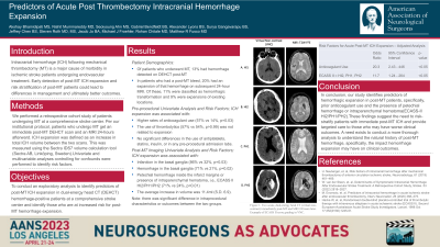
Introduction: Intracranial hemorrhage (ICH) following mechanical thrombectomy (MT) is a major cause of morbidity in ischemic stroke patients. Predictors of hemorrhage volume increase are not well-known. We seek to identify factors associated with ICH increase on imaging evaluation in post-MT patients.
Methods: We conducted a retrospective cohort study of 394 patients undergoing MT at a comprehensive stroke center between 2018 and 2021. Per protocol, patients in our cohort had dual-energy head CT (DEHCT) immediately post-MT and MRI 24 hours afterwards. Of these patients, 35 (9%) had ICH detected on their DEHCT scan. An increase in ICH was defined as an increase in blood volume between the two scans. Demographics, NIH stroke scale, PMH, medications, thrombolytic use, hemorrhage location (using ECASS II scores), number of passes, and recanalization score were collected. We conducted initial univariate analysis for hemorrhage increase to identify risk factors and performed multivariate analysis using logistic regression with covariates that had p< 0.10. Our adjusted analysis used p< 0.05 for statistical significance.
Results: Increase in ICH was seen in 7/35 patients on the 24-hour MRI. Of these, 4 were classified as hemorrhagic transformation and 3 were expansions of existing locations. Univariate analysis showed that anticoagulant use (57% vs 14%, p=0.03) and petechial hemorrhage inside the infarct margins or presence of intraparenchymal hematoma (ECASS II score HI2, PH1, PH2) (71% vs 14%, p< 0.01) on DEHCT were associated with hemorrhage increase. Multivariate regression was then performed for ECASS II scores and anticoagulation. Odds ratios were 8.00 (p < 0.05) for anticoagulant use and 15.00 (p < 0.05) for ECASS II HT2, PH1, or PH2 scores in predicting ICH increase.
Conclusion : Expansion or new hemorrhage on 24-hour MRI compared to immediate post-thrombectomy DECT is significantly associated with prior anticoagulant use and petechial hemorrhage inside the infarct margins (HT2) or presence of intraparenchymal hematoma (PH1 or PH2).
