Primary Stress and Strain Characterization of the Carpal Tunnel with Compression Ultrasound: A Novel Imaging Indicator
Friday, April 21, 2023


Sachin Govind (he/him/his)
Medical Student
Northwestern University Feinberg School of Medicine
Chicago, IL, US
ePoster Presenter(s)
Introduction: Pathologies affecting the carpal tunnel are easily visualized with ultrasound, but visualization and precise localization of the nerve entrapment is difficult to ascertain with traditional ultrasound imaging. Given the structural complexity of the carpal tunnel and its surrounding tissues, work is required to more concretely image and characterize the dynamic strains on the median nerve with nerve entrapment and after surgical repair.
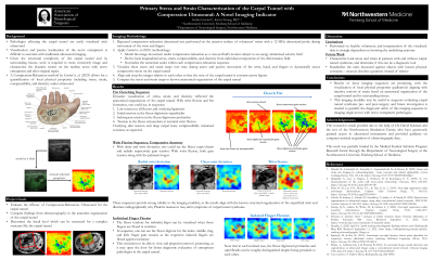
Methods: Repeated compression-relaxation ultrasound was performed on the anterior surface of volunteers’ wrists with a 12 MHz ultrasound probe during movement of the wrist and fingers. By modeling image movement under compression-relaxation as a viscous fluid’s motion subject to an energy-minimized velocity field, measures of local longitudinal stress, strain, compressibility, and elasticity were derived from components of the deformation field. Visualization of these stress and strain maps over time during active and passive movement was performed to dynamically assess compressive stress on the carpal tunnel.
Results: Dynamic visualization of stress, strain, and elasticity reflected the anatomical organization of the carpal tunnel. With wrist flexion and fist formation, one could see the flexor digitorum superficialis and profundus gain tension initially, with overlying skin remaining under low tension, and finally the flexor retinaculum gaining tension at terminal wrist flexion. This sequence provides validity to the imaging modality, as the results align with the structural organization of the superficial wrist and illustrate radiographically why Phalen’s maneuver may elicit symptoms of carpal tunnel syndrome.
Conclusion : Results of these imaging sequences are promising, with the visualization of local physical properties qualitatively aligning with intuitive notions of strain based on anatomical organization of the carpal tunnel and its surrounding tissues. This imaging modality may be useful to surgeons evaluating carpal tunnel syndrome pre- and post-surgery, and future investigation is needed to quantify the diagnostic utility of this imaging sequence in imaging single nerves with nerve entrapment pathologies.
