Exposed surgical area by the endoscope vs the microscope
Exposed Surgical Area by the Endoscope vs the Microscope
Friday, April 21, 2023


Esteban Quiceno Restrepo, MD (he/him/his)
Research Fellow
Banner University Hospital
Phoenix, Arizona, United States
ePoster Presenter(s)
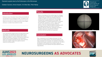
Introduction: Endoscopic access to the posterior fossa provides several benefits over the microscope but can be challenging to master. The objective of this experiment was to quantify the difference of surgical area of exposure between the microscope, the 0-degree endoscope and the 30-degree endoscope, in a posterior fossa simulation box model.
Methods: A box model with the average volume of an adult posterior fossa was designed. The viewable area with the 0-degree and 30-degree endoscopes set at various distances from a defined target were compared. In part two of the study the maximal area exposed with the maximal angulation of the microscope, 0-degree and 30-degree endoscopes through various craniotomy sizes were compared. Images were captured using the respective recording systems.
Results: Based on fixed distances from a defined target, the 30-degree endoscope had on average 40% more viewable area than the 0-degree endoscope. At 5mm the 30-degree endoscope provided up to 68% more viewable area. This advantage decreased with increasing distance from the target. With maximal angulation, the maximal exposed area on one side of the model with a craniotomy of 1.0cm2, 1.5cm2, 2.0cm2, 2.5cm2, 3.0cm2 with the microscope was 3859mm2, 4901mm2, 5541mm2, 5808mm2, 5944mm2 respectively, with the 0-degree endoscope it was 4778mm2, 5541mm2, 6082mm2, 6361mm2, 6361mm2, respectively and with the 30-degree endoscope it was 5410mm2, 5816mm2, 6361mm2, 6361mm2 and 6361mm2 respectively.
Conclusion : When comparing the 0 and 30-degree endoscopes, the latter achieves more surgical exposure with decreasing distance to the target. Compared to the microscope, the endoscope enhances the viewable area through a craniotomy of a particular size. This effect is less apparent as the craniotomy size increases. Angled-lens endoscopes use must be balanced with surgical experience due to image distortion.
