Spatiotemporal mapping and decoding of oculomotor control in human frontal eye fields
Spatiotemporal Mapping and Decoding of Oculomotor Control in Human Frontal Eye Fields
Friday, April 21, 2023


Stephano Chang, MD, PhD
Neurosurgery Resident
University of British Columbia
Vancouver, British Columbia, Canada
ePoster Presenter(s)
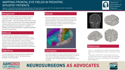
Introduction: The frontal eye fields (FEFs) are linked to oculomotor control and hypothesized to reside in the prefrontal cortex, where electrical stimulation reportedly evokes contraversive eye movements. The exact location and function of the FEFs in humans is controversial with studies implicating multiple putative regions, including the posterior middle frontal gyrus and the inferior precentral gyrus. Additionally, these studies are performed in the operating room, where stimulation localization is suboptimal and neural function may be influenced by anesthetics. Stereo-electroencephalography (SEEG) is a minimally invasive technique for epilepsy work up, using depth electrodes in the brain. It provides a unique opportunity to collect human neurophysiological data outside of the operating room. The objective of this study was to identify and map the FEFs in pediatric and adult patients after SEEG implantation in the post-operative, anesthestic-free setting.
Methods: Five pediatric and five adult subjects undergoing non-lesional epilepsy workup were enrolled into this prospective, IRB-approved study, and received brain MRI prior to SEEG implantation, followed by post-operative CT head for precise electrode localization. Postoperative stimulation testing and SEEG recordings were performed along with time-aligned video of the subjects’ eyes while performing gaze-related tasks.
Results: Stimulation testing elicited contraversive head turning with or without eye deviation, depending on the site of stimulation. Low-threshold sites eliciting these stereotyped movements were located just deep to the inferior precentral gyrus. Stimulation of sites in the posterior middle frontal gyrus did not elicit eye deviation movements in our subjects in the post-operative setting. Analysis of SEEG from electrodes in the inferior precentral gyrus revealed high correlations to eye movements.
Conclusion : Our findings suggest that the human FEFs are located more posteriorly than widely held and reported in textbooks, involving the motor cortex. Further testing in pediatric and adult subjects is warranted to confirm this hypothesis and test for differences in these populations.
