Application of Radiomics to the Differential Diagnosis of Temporal Bone Skull Base Lesions: A Pilot Study
Friday, April 21, 2023


Matt Findlay, BS
Medical Student
University of Utah School of Medicine
Salt Lake City, Utah, United States
ePoster Presenter(s)
Introduction: Temporal bone skull base pathologies represent a complex differential with surgical resection in this location often having significant morbidity. Radiomics involves the use of mathematical quantification of imaging data beyond simple intensity, size, and location to inform diagnosis and prognosis. We sought to assess the feasibility of using radiomic parameters to help predict temporal bone tumor type.
Methods: A total of 117 radiomic parameters (Slicer, Pyradiomics) were analyzed in each of 5 MRI sequences (T1 without contrast, T1 with contrast, T2, FLAIR, ADC). Statistical analysis was used to delineate known primary, metastatic/secondary and lymphoma lesions using radiomics.
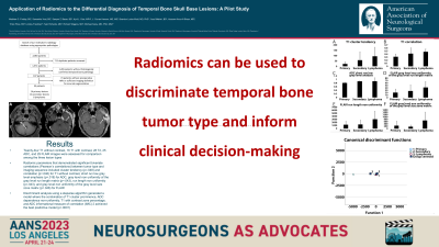
Results: A total of 14 primary, 12 secondary, and 8 lymphoma lesions were assessed. Mean tumor volumes of 2.98±2.11, 3.28±2.31, and 12.16±7.1 cm3 were seen for primary, secondary and lymphoma tumors, respectively (p=0.2, ANOVA). No significant difference in mean intensity values for any sequence helped distinguish tumors (p>0.05, ANOVA). Cluster tendency (p=0.05), correlation (p=0.05), short run low gray level emphasis (p=0.02), gray level non-uniformity of both the run length matrix (p=0.04) and size zone matrix (p=0.05), and run length non-uniformity (p=0.04) were significantly correlated with diagnostic accuracy (Pearson’s correlation). Discriminant analysis using a stepwise algorithm generated a model where T1 cluster prominence, ADC dependence non-uniformity, T1 with contrast zone percentage, and ADC Informational Measure of Correlation (IMC) 2 achieved the best predictive model (p=0.0001).
Conclusion : While this pilot study requires further testing and validation, these data support the exploration of radiomics in temporal bone radiographic diagnostics.
