Endovascular management of cranial neurovascular trauma.
Endovascular Management of Cranial Neurovascular Trauma
Friday, April 21, 2023


David C. Lauzier, n/a
Medical Student
Washington University School of Medicine
St. Louis, Missouri, United States
ePoster Presenter(s)
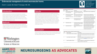
Introduction: Traumatic vascular injuries to the head and neck following blunt or penetrating trauma are common and carry a risk of severe disability or even death. Streamlined screening, diagnosis, and treatment of these injuries is key to improve patient outcomes. Advances in imaging continue to improve the accuracy of non-invasive diagnosis of these injuries while new clinical data provide better evidence for optimal management, whether medical or invasive.
Methods: Here, we reviewed the current literature to assess contemporary indications and management strategies for cranial neurovascular trauma. This included presentation modalities, diagnostic workup (both imaging and non-imaging), current classification, non-invasive management, and interventional management.
Results: Presentation of cranial neurovascular trauma can be classified as either blunt or penetrating, which each carry unique risks of vascular injury. Clinically, blunt injuries appear to be underrecognized while penetrating injuries are more frequently apparent. Catheter angiography traditionally was used as a first line for imaging of these lesions, but has fallen out of favor with CTA and MRA frequently employed. Catheter angiography continues to carry the advantage of serving as a vehicle for immediate endovascular treatment. Treatment indications for blunt and penetrating injuries followed the Biffl classification or the Roon and Christensen classification, respectively. In all cases, a thoughtful multidisciplinary approach is necessary to optimize patient outcomes.
Conclusion : Cranial neurovascular trauma is common and can lead to poor patient prognosis. Recent evolutions in imaging techniques and alignment of modern neurointerventional methods with traditional grading scales has led to improved patient outcomes.
