Morphological assessment of whole Spine utilizing Computed Tomography versus MRI based synthetic Computed Tomography
Morphological Assessment of Whole Spine Utilizing Computed Tomography versus MRI Based Synthetic Computed Tomography
Friday, April 21, 2023


Daniel Davidar, MBBS
Research Fellow
Johns Hopkins School of Medicine
Baltimore, Maryland, United States
ePoster Presenter(s)
Introduction: Reconstruction of a ‘synthetic Computed Tomography’(sCT) derived from a Magnetic Resonance Imaging (MRI) scan has been successfully performed for the hip
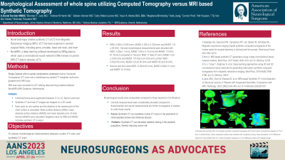
Methods: A female cadaver with no known spinal complications underwent a routine Computed Tomography (CT) scan and an MRI scan in a 3T Siemens with specific sCT protocols. sCT images were generated from three-dimensional radiofrequency-spoiled T1-weighted multi-echo gradient-echo MR images using a commercially available deep learning-enabled software BoneMRI (MRI Guidance, Netherlands). Individual bones were segmented between C1 to L5, Sacrum and ilium. Synthetic CT and real CT images are mapped on a 3D model. Each point on one surface and the distance to the nearest point of the other surface is calculated. Mean surface distance (MSD), mean absolute surface distance (MASD) and mean absolute error of radiodensity (MAER) were calculated. Negative value for MSD and MASD indicates synthetic CT is larger.
Results: MSD: 0.008 ± 0.209 (mm), MASD: 0.355 ± 0.128 (mm) and MAER: 114 ± 28 (HU). Cervical morphological measurements were elevated with MSD: -0.26±1.7 (mm), MASD: 0.49 ± 0.14 (mm) and MAER: 173.57± 61.76 (HU) compared to Thoracic: MSD: 0.19±0.07 (mm), MASD: 0.35 ± 0.06 (mm) and MAER: 107.83± 6.24 (HU) and Lumbar: MDS -0.02±0.05 (mm), MASD 0.21±0.05 (mm) and MAER 90.2±8.34 (HU). Sacrum and ilium were MSD: -0.02±0.05 (mm), MASD 0.29±0.01 (mm) and MAER 14.19 (HU).
Conclusion : Morphological results were comparable compared to those reported in the literature. Cervical measurements were considerable elevated compared to thoracolumbar and sacral measurements but further investigation is needed to verify these results.
