Four-dimensional brain atlas of executive control
Four-dimensional Brain Atlas of Executive Control
Friday, April 21, 2023


Matthew T. Brennan (he/him/his)
Medical Student
Wayne State University
Detroit, Michigan, United States
ePoster Presenter(s)
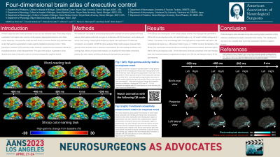
Introduction: Humans utilize executive control processes to carry out non-automatic tasks. These tasks require coordination from higher brain centers to both suppress inappropriate behaviors and initiate correct responses. The prefrontal cortex has been shown in previous studies using individual data to elicit high-gamma augmentation in tasks requiring heightened executive control. In fact, following resection of prefrontal regions, patients have shown decline in executive control function. The goal of this study is to generate a novel, dynamic brain atlas to minimize postoperative cognitive decline.
Methods: We studied 547 non-epileptic intracranial electrode sites sampled from seven patients with focal epilepsy. Each patient performed two types of verbal tasks with 40 stimuli each: word-reading and Stroop color-naming. Following time-frequency analysis, each participant’s 3-D MRI was converted to a normalized 3-D MRI for group analysis. Mixed model analysis compared high-gamma cortical activation prior to response onset between the word-reading and Stroop color-naming tasks. Based on the mixed model analysis, we visualized the white matter connectivity between the brain regions exhibiting simultaneous high-gamma augmentation.
Results: In the Stroop color-naming task, mixed model analysis showed more high-gamma augmentation 600 to 400 ms pre-response onset in the prefrontal region (e.g., left caudal middle-frontal gyrus; p = 0.0054). Conversely, in the word-reading tasks, more high-gamma augmentation was seen in the occipitotemporal region (e.g., left posterior fusiform gyrus; p = 0.0002). Dynamic tractography in the Stroop color-naming task showed functional connectivity enhancement between prefrontal regions 500 to 400 ms pre-response onset. On the other hand, functional connectivity in the word-reading tasks was enhanced between occipitotemporal regions from 500 ms pre-response onset to 50 ms post-response.
Conclusion : Prefrontal regions were activated during tasks requiring higher executive control, whereas occipitotemporal regions supported word reading. The resulting atlas may serve as a valuable asset to prevent postoperative decline in cognitive function.
