Early and Long-Lasting White Matter Damage in The Perforant Pathway Following Subarachnoid Hemorrhage In Mice
Early and Long-lasting White Matter Damage in the Perforant Pathway Following Subarachnoid Hemorrhage in Mice
Friday, April 21, 2023

.jpg)
Gavin Britz, MD, MBA, MPH (he/him/his)
Department Chair
Houston Methodist Hospital
Houston, TX, US
ePoster Presenter(s)
Introduction: Subarachnoid hemorrhage (SAH) is when the blood extravasates into the subarachnoid space. SAH is the least frequent stroke but has a high fatality rate (40%) and often debilitates people of working age (under 55) and impedes them from going back to work. In our mouse model of SAH, we observed the development of chronic behavioral abnormalities consistent with those observed in humans. Our RNA next-generation sequencing study of the hippocampus, structure of learning and memory, at 4-days, showed a significant downregulation in the myelin/oligodendrocytes-related genes. We leveraged these observations and proposed that oligodendrocytes and white matter damages occur in the perforant pathway, connecting the entorhinal cortex and the hippocampus, early on, and play a major role in cognitive deficits observed.
Methods: SAH was induced by the endovascular perforation of the circle of Willis. In one group, brain tissues were processed for immunohistochemistry (IHC) of MBP (Myelin Basic Protein) at 24h and 96h. In the second group, we conducted diffusion tensor magnetic resonance imaging in Magnetic Resonance Imaging (DTI-MRI) in mice 10-12months following SAH to assess the water diffusion of the brain fibers and reflect their structure.
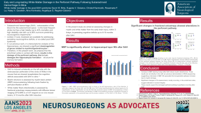
Results: At 24h and 96h, IHC for MBP is increased in all layers of the hippocampus compared to Sham animals contradicting our transcriptome analysis. This suggests that destruction of myelin may uncover more epitopes explain an increase in staining. Increase in fractional anisotropy, measuring the directionality of the white fiber, was observed in the entorhinal cortex but no significant change was observed in the hippocampus, suggesting a remodeling of the white fibers over time.
Conclusion : While further investigations are needed, the present results confirm early time points and long- lasting white matter abnormalities occurring after SAH.
