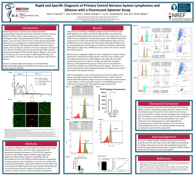
Rapid and Specific Diagnosis of Primary Central Nervous System Lymphoma and Glioma with a Fluorescent Aptamer Assay
Friday, April 21, 2023

.jpg)
Dara S. Farhadi, MD (he/him/his)
Research Fellow
University of Arizona College of Medicine - Phoenix
Phoenix, Arizona, United States
ePoster Presenter(s)
Introduction: Brain tumor diagnostics rely on immunohistochemistry (IHC), However, IHC is too slow to provide specific intraoperative diagnoses. This potentially impacts patient care. Aptamers are oligonucleotide probes engineered to label tissue similarly to IHC, but within an intraoperative timeframe. We aim to develop diagnostic aptamers to intraoperatively differentiate high-grade glioma (HGG) from primary central nervous system lymphoma (PCNSL).
Methods: Fluorescent aptamers specific to CD20 (PCNSL target) and GFAP (HGG target) were engineered utilizing SELEX. We optimized staining parameters to label PCNSL and HGG cell lines from 5 to 60 minutes and aptamer concentrations between 200nM to 1600nM. Welch’s t-test was used for statistical comparisons.
Results: Via flow cytometry, 95% of PCNSL cells stained with CD20 aptamer and 95% of HGG cells stained with GFAP aptamer. However, we also found 98.5% of PCNSL cells were stained with GFAP aptamer and 37.3% of HGG cells stained with CD20 aptamer. The median fluorescence of a GFAP aptamer with HGG cells was 6,286 relative fluorescence units (RFUs) vs PCNSL cells 887 RFUs (p < 0.001). However, CD20 aptamer fluorescence of HGG cells was not significant, 10,814 RFUs vs PCNSL cells 3,893 RFUs (p=0.10). Optimal aptamer concentration was 400nM and optimal staining time was 20 minutes.
Conclusion : GFAP aptamers strongly bound glioma cells but unexpectedly had high binding affinity to PCNSL cell lines. We also found CD20 aptamers unexpectedly bound glioma cell lines, though not significant. This may indicate confounding factors such as non-specific binding or improper annealing that warrant further exploration.
