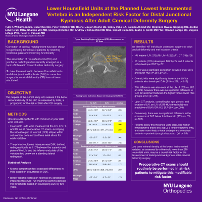
Lower Hounsfield Units at the Planned Lowest Instrumented Vertebra is an Independent Risk Factor for Distal Junctional Kyphosis After Adult Cervical Deformity Surgery
Lower Hounsfield Units at the Planned Lowest Instrumented Vertebra Is an Independent Risk Factor for Distal Junctional Kyphosis After Adult Cervical Deformity Surgery
Friday, April 21, 2023


Peter G. Passias, MD
Spine Surgeon
NYU School of Medicine
New Canaan, CT, US
ePoster Presenter(s)
Introduction: The association of Hounsfield units(HU) and junctional pathologies has emerged as a topic of interest in thoracolumbar surgery. The relationships between HUs and complications like distal junctional kyphosis(DJK) in cervical deformity(CD) surgery have not been elucidated. We sought to determine if the bone mineral density of the LIV, as assessed by HUs, is prognostic for the risk of DJK after CD surgery
Methods: CD patients with 2-year(2Y) data included. HUs were measured at the LIV, LIV+1, C3 and C7 on all preoperative CT scans, averaging the widest region of interest(ROI) ellipse within sub-cortical bone across three axial slices for each vertebra. The primary outcome measure was complications, including DJK, defined radiographically as ≥10° change between the end plates of a vertebra and vertebra+2. Means comparison test assessed differences in HUs based on occurrence of complications, linear regression assessed correlation of HUs with risk factors, and multivariable logistic regression followed by conditional inference tree(CIT) run machine learning derived HU thresholds based on developing complications.
Results: Included: 107 CD patients. HU means- LIV: 272±79, LIV+1: 252±71, C3: 338±109, C7: 294±97. 20.6% developed DJK and 6.3% developed DJF by 2Y. There was a significant correlation between lower LIVs and lower HUs (r=.351, p=.01), as well as age and HUs at the LIV(r=.364,p < .001). Age and HUs did not correlate with change in the DJK angle(both p>.1). HUs were lower at the LIV for patients who developed DJK(219 vs 286,p=.018). Upon CIT analysis, an LIV threshold of 245 HUs was predictive of DJK(OR: 5.0,[1.2-17.7]; p=.023).
Conclusion : Low bone mineral density at the lowest instrumented vertebra, as assessed by a threshold lower than 245 Hounsfield units, may be a crucial risk factor for the development of distal junctional kyphosis after cervical deformity surgery. Preoperative CT scans should routinely be performed in at-risk patients to mitigate this modifiable risk factor.
